
CORNEAL SUBBASAL NERVE PLEXUS EVALUATION BY IN VIVO CONFOCAL MICROSCOPY IN MULTIPLE SCLEROSIS
DOI:
https://doi.org/10.48560/rspo.15222Resumo
PURPOSE
Multiple Sclerosis (MS) is the most frequent cause of neurologic disability in young adults, characterized by demyelination and axonal degeneration. Monitoring this last component remains an important challenge. Our study aims to access corneal subbasal nerve plexus morphology by in vivo confocal microscopy (CCM) and explore the possibility of using this noninvasive technology to obtain a biomarker of axonal degeneration.
METHODS
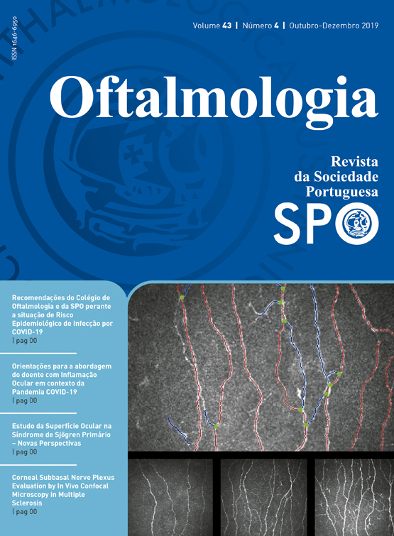
In this cross-sectional study 30 patients with MS and 22 healthy controls were included. All participants underwent full ophthalmology standard evaluation, CCM and optical coherence tomography (OCT). The following corneal subbasal nerve plexus morphology parameters were analysed: corneal nerve fibre density (CNFD), corneal nerve branch density (CNBD), corneal nerve fibre length (CNFL) and corneal nerve fibre tortuosity (CNFT). Neurological disability of MS patients was accessed using Expanded Disability Status Scale (EDSS) and MS Severity Score (MSSS).
RESULTS
Compared to controls, MS patients have lower CNFD, CNBD and CNFL (p<0.001) but no significant difference was found related to CNFT (p=0.108). No significant differences were found related to corneal subbasal plexus parameters between MS patients with or without optic neuritis (MSON vs MSNON). CNFD and temporal-inferior peripapillary retinal nerve fibre layer (ppRNFL) showed inverse association both with EDSS (rS=-0.62, p<0.001 for CNFD; rS=-0.53, p=0.003 for temporal-inferior ppRNFL) and MSSS (rS=-0.44, p=0.018 for CNFD; rS=-0.49, p=0.009 for temporal-inferior ppRNFL) scores.
CONCLUSIONS
CNFD, CNBD and CNFL are decreased in MS patients, suggesting axonal degeneration. Further longitudinal studies are needed to confirm whether CNFD could be a promising imaging parameter in MS severity evaluation.
Keywords: multiple sclerosis, axonal degeneration, optical coherence tomography, corneal confocal microscopy, expanded disability status scale.
Downloads
Downloads
Publicado
Como Citar
Edição
Secção
Licença
Não se esqueça de fazer o download do ficheiro da Declaração de Responsabilidade Autoral e Autorização para Publicação e de Conflito de Interesses
O artigo apenas poderá ser submetido com esse dois documentos.
Para obter o ficheiro da Declaração de Responsabilidade Autoral, clique aqui
Para obter o ficheiro de Conflito de Interesses, clique aqui

