Características de Imagem Multimodal da Síndrome de Múltiplos Pontos Brancos Evanescentes
DOI:
https://doi.org/10.48560/rspo.25563Palavras-chave:
Imagem Multimodal, Síndrome dos Pontos Brancos/diagnósticoResumo
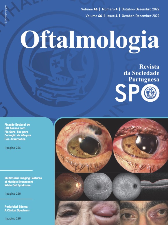
A 24-year-old male patient with no medical and ophthalmological background presented to our Ophthalmology Department with complaints of relative paracentral scotoma in the right eye (OD) and decreased visual acuity (VA) since awakening. The distance-corrected VA of the OD was 20/100. Biomicroscopy was uneventful, and intraocular pressure was 12 mmHg. Ophthalmoscopy showed multiple small gray-white spots in the posterior pole of the OD, documented by color fundus photography (a). Fluorescein angiography revealed multiple clustered hyperfluorescent dots in a “wreath-like” pattern that matched the clinically observed spots (b). Indocyanine green angiography showed multiple hypocyanecent spots, in a greater number than those observed in the fundus or AF (c). Macular optic coherence tomography revealed disruption of the outer retinal layers (d).
The diagnosis of multiple evanescent white dot syndrome (MEWDS) was assumed, and the patient was periodically observed. After four weeks, there was a complete resolution of the symptoms. VA was 10/10, and the previously observed changes faded away completely.
MEWDS is one of the diagnoses within the family of white dot syndromes, first described by Jampol LM and colleagues.1 The white dot syndromes constitute a group of inflammatory chorioretinopathies in which the common defining clinical feature is the presence of multiple, discrete, white lesions located at the deeper levels of the retina and choroid primarily found in young adults. Both infectious and noninfectious entities are considered in the differential diagnosis of the white dot syndromes. The noninfectious diseases include sarcoidosis, Vogt-Koyanagi-Harada syndrome, sympathetic ophthalmia, and intraocular lymphoma. Syphilis, tuberculosis, and diffuse unilateral subacute neuroretinitis are infectious entities that should also be considered in the differential diagnosis.2 Symptoms of MEWDS include unilateral blurred vision, visual field loss, photopsia, and floaters. The symptoms and fundus findings usually improve in 2-6 weeks without treatment.1
Downloads
Referências
Jampol LM, Sieving PA, Pugh D, Fishman GA, Gilbert H. Multiple evanescent white dot syndrome. I. Clinical findings. Arch Ophthalmol. 1984;102:671-4.
Mount GR, Kaufman EJ. White Dot Syndromes. Treasure Island; StatPearls: 2022.
Downloads
Publicado
Como Citar
Edição
Secção
Licença
Direitos de Autor (c) 2022 Revista Sociedade Portuguesa de Oftalmologia

Este trabalho encontra-se publicado com a Creative Commons Atribuição-NãoComercial 4.0.
Não se esqueça de fazer o download do ficheiro da Declaração de Responsabilidade Autoral e Autorização para Publicação e de Conflito de Interesses
O artigo apenas poderá ser submetido com esse dois documentos.
Para obter o ficheiro da Declaração de Responsabilidade Autoral, clique aqui
Para obter o ficheiro de Conflito de Interesses, clique aqui


