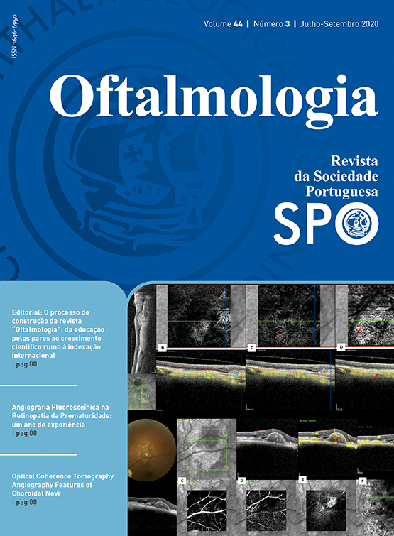
Optical coherence tomography angiography features of choroidal nevi
DOI:
https://doi.org/10.48560/rspo.19806Abstract
Purpose: To describe the imaging features of choroidal nevi using optical coherence tomography angiography (OCT-A) and optical coherence tomography with enhanced depth imaging (EDI-OCT).
Material and Methods: Retrospective observational case series of patients with choroidal nevi. The tumor and overlying retina structural features were analyzed with EDI-OCT. The OCT-A images were evaluated for vascular changes at the level of the retinal plexus, outer retina, choriocapillaris and choroid. Statistical analysis were performed using IBM SPSS Statistics.
Results and Discussion: 21 patients, 73% female, were included. The mean age was 55±10 years old. The mean tumor thickness was 359±42 µm and the mean largest basal diameter was 3089±261µm. 27% of the nevi had their epicenter at the macula. US showed a solid flat lesion with high reflectivity. With EDI-OCT all lesions appeared as a highly reflective band within the choriocapillaris with posterior shadowing. 67% of the nevi had drusen and 25% had drusenoid pigment epithelium detachment (dPED). Chronic changes in the retina were found in 52% and subretinal fluid in 16%. On OCT-A the superficial and deep capillary plexus were normal. A neovascular membrane was detected in 1 case and a polypoidal vasculopathy in 3 cases. At the level of the choriocapillaris, 16% were hyperreflective, 74% were isorreflective and 10% were hyporreflective.
Conclusion: Our work demonstrated that OCT-A may be useful in the diagnosis of vascular complications associated to small stable nevi. The identification of vascularized PEDs is crucial in order to decide treatment, avoiding further visual impairment.
Downloads
Downloads
Published
How to Cite
Issue
Section
License
Do not forget to download the Authorship responsibility statement/Authorization for Publication and Conflict of Interest.
The article can only be submitted with these two documents.
To obtain the Authorship responsibility statement/Authorization for Publication file, click here.
To obtain the Conflict of Interest file (ICMJE template), click here


