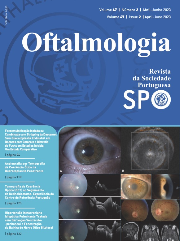
Optical Coherence Tomography Angiography in Penetrating Keratoplasty
DOI:
https://doi.org/10.48560/rspo.28278Keywords:
Corneal Neovascularization, Fluorescein Angiography, Keratoplasty, Penetrating, Tomography, Optical CoherenceAbstract
INTRODUCTION: Penetrating keratoplasty (PK) is one of the most common surgical procedure in corneal transplantation worldwide. Graft failure and rejection risk progressively increases with the increasing number of quadrants with corneal neovascularization (CNV). Optical coherence tomography angiography (OCTA) is a noninvasive imaging technique that has been widely used to visualize vascular abnormalities in the retina and some studies have shown its potential use in the anterior segment (AS) of the eye. The purpose of this study was to investigate the potential of OCTA technology to image and describe quantitatively CNV in eyes submitted to PK.
MATERIAL AND METHODS: A cross-sectional study was performed, including 20 eyes from 18 patients submitted to PK at least 5 years before and with no history of graft rejection. All eyes underwent anterior segment slit-lamp photography (SLP) and OCTA with en face, b-scans and c-scans imaging. The vessel density (VD) was analyzed in the inferior, nasal and temporal corneal margin in all patients. The measurements were calculated after binarization with ImageJsoftware, using OCTA scans with 6 × 6 mm in a depth of 800 μm.
RESULTS: The mean age was 59 years-old and most patients were submitted to PK due to corneal leucoma, followed by keratoconus, and a few had Fuchs endothelial corneal dystrophy and bullous keratopathy. The mean total VD was 50.16% and it was higher in the temporal quadrant and lower in the inferior one. However, there were no statistically significant differences between the 3 analyzed areas (p=0.801) or between each area and the other two and there was nocorrelation between the areas. OCTA was able to identify abnormal vessels when SLP apparently showed no abnormal vessels; OCTA was able to distinguish between larger and smaller vessels; OCTA scans allowed the investigation of several corneal planes.
CONCLUSION: OCTA can become a new method for monitoring corneal diseases. It may allow the qualitative and quantitative follow-up of patients submitted to PK over time and to detect the appearance of CNV earlier than through SLP. OCTA applied to the anterior segment has promising and valuable features.
Downloads
References
Mathews PM, Lindsley K, Aldave AJ, Akpek EK. Etiology of global corneal blindness and current practices of corneal transplantation: A focused review. Cornea. 2018;37:1198-203. doi: 10.1097/ICO.0000000000001666
An DT, Dart JK, Holland EJ, Kinoshita S. Corneal transplantation. Lancet. 2012;379:1749-61. doi: 10.1016/S0140- 6736(12)60437-1
Bachmann B, Taylor RS, Cursiefen C. Corneal neovascularization as a risk factor for graft failure and rejection after kera- toplasty: an evidence-based meta-analysis. Ophthalmology. 2010;117:1300-5.e7. doi: 10.1016/j.ophtha.2010.01.039.
Bir R, Marmalidou A, Amouzegar A, Chen Y, Dana R. Animal models of high-risk corneal transplantation: A compre- hensive review. Exp Eye Res. 2020;198:108152. doi: 10.1016/j. exer.2020.108152
Sugar J. The Collaborative Corneal Transplantation Studies. Arch Ophthalmol. 1992;110:1392-403. doi: 10.1001/ar- chopht.1992.01080230015004
Stanzel TP, Devarajan K, Lwin NC, Yam GH. Comparison of optical coherence tomography angiography to indocyanine green angiography and slit lamp photography for corneal vascularization in an animal model. Sci Rep. 2017;2018:1–11. doi: 10.1038/s41598- 018- 29752-5
Cai Y, Alio JL, Wilkins MR, Ang M. Serial optical coherence tomography angiography for corneal vascularization. Graefe’s Arch Clin Exp Ophthalmol. 2016. doi: 10.1007/s00417-016- 3505-9
Chan SY, Pan CT, Feng Y. Localization of corneal neovascularization using optical coherence tomography angiography. Cornea. 2019;38:888–95. doi: 10.1097/ICO.0000000000001931
Almeida I, Dias L, Jesus J, Fonseca I, Matias MJ, Pedro JC. Optical coherence tomography angiography in herpetic leucoma. BMC Medical Imaging. 2022; 22:17. doi: 10.1186/s12880-022-00747-z.
Jesus J, Dias L, Almeida I, Costa T, Chibante-Pedro J. analysis
of conjuntival vascular density in scleral contact lens wearers using optical coherence tomography angiography. Contact Lens Anterior Eye. 2020; 45:1. doi: 10.1016/j.clae.2020.12.066
Patel CN, Antony AK, Kommula H, Shah S, Singh V, Basu S. Optical coherence tomography angiography of perilim- bal vasculature: validation of a standardised imaging algorithm. Br J Ophthalmol. 2019;0:1–6. doi: 10.1136/bjophthalmol-2019-314030.
Schneider CA, Rasband WS, Eliceiri KW. NIH Image to ImageJ: 25 years of image analysis. Nat Methods. 2012;9:671–5. doi: 10.1038/nmeth.2089.
Nanji A, Redd T, Chamberlain W, Schallhorn JM, Chen S, Ploner S, et al. Application of Corneal Optical Coherence Tomography Angiography for Assessment of Vessel Depth in Corneal Neovascularization. Cornea. 2020;39:598-604. doi: 10.1097/ICO.0000000000002232
Ang M, Sim DA, Keane PA, et al. Optical coherence tomography angiography for anterior segment vasculature imag- ing. Ophthalmology. 2015;122:1740–7. doi: 10.1016/j.Oph- tha.2015.05.017
Li S, Li L, Zhou Q, Gao H, Liu M, Shi W. Blood vessels and lymphatic vessels in the cornea and iris after pen- etrating keratoplasty. Cornea. 2019;38:742-7. doi: 10.1097/ ICO.0000000000001922
Cai Y, Alio JL, Wilkins MR, Ang M. Serial optical coherence tomography angiography for corneal vascularization. Graef- es Arch Clin Exp Ophthalmol. 2017;255:135-9. doi: 10.1007/ s00417-016-3505-9
Ang M, Tan AC, Ming C, Cheung G, Keane PA, Dolz-Marco R. Optical coherence tomography angiography: a review of cur- rent and future clinical applications. Graefes Arch Clin Exp Ophthalmol. 2018;256:237–45. doi: 10.1007/s00417-017-3896-2
Binotti WW, Mills H, Nosé RM, Wu HK, Duker JS, Hamrah P. Anterior segment optical coherence tomography angiog- raphy in the assessment of ocular surface lesions. Ocul Surf. 2021; 22:86-93. doi: 10.1016/j.jtos.2021.07.009
Luo M, Li Y, Zhuo Y. Advances and current clinical applications of anterior segment optical coherence tomogra- phy angiography. Front Med. 2021;8:721442. doi: 10.3389/ fmed.2021.721442.
Siddiqui Y, Yin J. Anterior segment applications of optical coherence tomography angiography. Semin Ophthalmol. 2019; 34: 264-9. doi: 10.1080/08820538.2019.1620805
Akagi T, Uji A, Okamoto Y, Suda K, Kameda T, Nakanishi H, et al. Anterior Segment Optical Coherence Tomography Angiography Imaging of Conjunctiva and Intrasclera in Treated Primary Open-Angle Glaucoma. Am J Ophthalmol. 2019;208:313-22. doi: 10.1016/j.ajo.2019.05.008.
Downloads
Published
How to Cite
Issue
Section
License
Copyright (c) 2023 Revista Sociedade Portuguesa de Oftalmologia

This work is licensed under a Creative Commons Attribution-NonCommercial 4.0 International License.
Do not forget to download the Authorship responsibility statement/Authorization for Publication and Conflict of Interest.
The article can only be submitted with these two documents.
To obtain the Authorship responsibility statement/Authorization for Publication file, click here.
To obtain the Conflict of Interest file (ICMJE template), click here


