
Topography-Guided Corneal Crosslinking for Keratoconus: Is It Enough to Treat the Cone?
DOI:
https://doi.org/10.48560/rspo.28285Keywords:
Corneal Cross-Linking, Corneal Topography, Cross-Linking Reagents, Keratoconus, Patient Outcome AssessmentAbstract
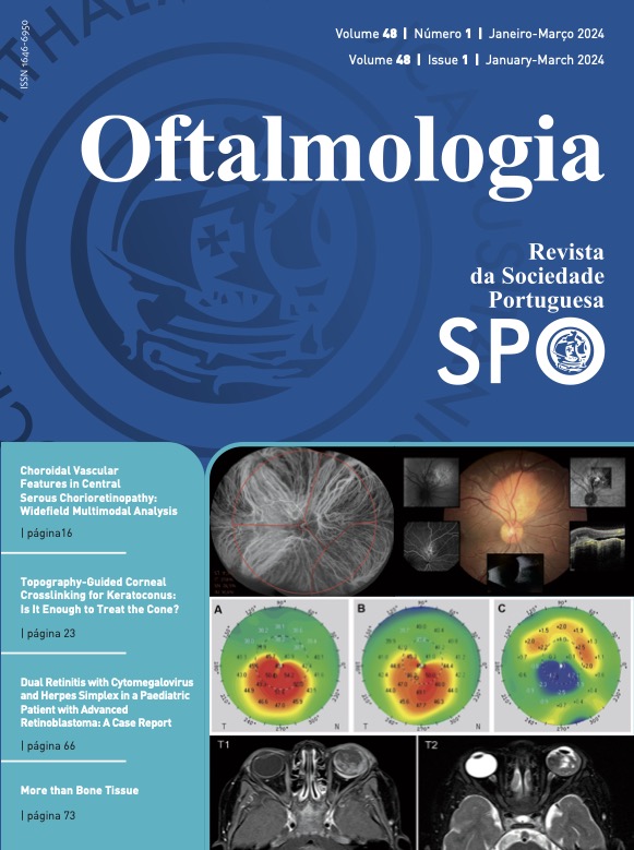
INTRODUCTION: Our objective is to report topographic and refractive outcomes in the first year following topography guided corneal crosslinking (TG-CXL) for keratoconus, with special focus given to the morphological changes in the superior, non-treated, corneal stroma.METHODS: Retrospective study. We included patients with progressive keratoconus who underwent TG-CXL: The epithelium was removed using phototherapeutic keratectomy with a 50 µm ablation within a 7.0 mm optic zone, followed by riboflavin application each 2 minutes for 10 minutes, and topography guided irradiation with ultraviolet-A, with treatment energies ranging from 10 to 5.4 J/cm2 and fluence of 10 mw/cm2. Patient data was collected at baseline, 3, 6 and 12 months postoperatively and included maximum keratometry (Kmax), mean anterior keratometry values within 5 points of a 3 mm diameter circle centered at the pupil, in the superior (S index) and inferior (I index) halves of the cornea, central corneal thickness, thinnest point pachymetry, subjective refraction and best corrected visual acuity (BCVA).
RESULTS: Twenty-seven eyes from 24 patients were included. Kmax was significantly flattened at 6 months (-0.92 ± 1.58 D; p=0.011) and 1 year (-0.83 ± 1.64 D; p=0.016) postoperatively. I index decreased significantly at 6 months (-0.81 ± 1.10 D; p=0.002) and 1 year (-0.83 ± 1.11 D; p=0.001), while the S index increased significantly at 6 months (0.93 ± 1.70 D; p=0.016) and 1 year (0.81 ± 1.42 D; p=0.008). There were no significant differences in Kmax (0.16 ± 0.99 D; p=0.457), I index (-0.004 ± 0.60 D; p=0.978) and S index (-0.04 ± 0.99 D; p=0.864) between 6 and 12 months postoperatively. The BCVA improved significantly at 1 year (difference of -0.13 ± 0.14 logMAR to baseline; p<0.001), while there was a significant myopic increase in spherical refractive error (difference of -1.05 ± 2.08 D to baseline; p=0.017).
CONCLUSION: TG-CXL leads to flattening of Kmax and superior corneal steepening at 1 year postoperatively. These keratometric changes seem to stabilize at 6 months. Spherical myopic refractive error increased and BCVA improved during follow-up. These results support TG-CXL as a valuable procedure in progressive keratoconus.
Downloads
References
Romero-Jiménez M, Santodomingo-Rubido J, Wolffsohn JS. Keratoconus: a review. Cont Lens Anterior Eye. 2010;33:157-66; quiz 205. doi:10.1016/j.clae.2010.04.006
Santodomingo-Rubido J, Carracedo G, Suzaki A, Villa-Collar C, Vincent SJ, Wolffsohn JS. Keratoconus: An updated review. Cont Lens Anterior Eye. 2022;45:101559. doi:10.1016/j.clae.2021.101559
Caporossi A, Mazzotta C, Baiocchi S, Caporossi T. Long-term results of riboflavin ultraviolet a corneal collagen cross-linking for keratoconus in Italy: the Siena eye cross study. Am J Ophthalmol. 2010;149:585-93. doi:10.1016/j.ajo.2009.10.021
Koller T, Mrochen M, Seiler T. Complication and failure rates after corneal crosslinking. J Cataract Refract Surg. 2009;35:1358-62. doi:10.1016/j.jcrs.2009.03.035
Asri D, Touboul D, Fournié P, Malet F, Garra C, Gallois A, et al. Corneal collagen crosslinking in progressive keratoconus: multicenter results from the French National Reference Center for Keratoconus. J Cataract Refract Surg. 2011;37:2137-43. doi:10.1016/j.jcrs.2011.08.026
Beckman KA, Gupta PK, Farid M, Berdahl JP, Yeu E, Ayres B, et al. Corneal crosslinking: Current protocols and clinical approach. J Cataract Refract Surg. 2019;45:1670-9. doi:10.1016/j.jcrs.2019.06.027
Wu D, Lim DK, Lim BX, Wong N, Hafezi F, Manotosh R, et al. Corneal cross-linking: the evolution of treatment for corneal diseases. Front Pharmacol. 2021;12:686630. doi:10.3389/fphar.2021.686630
Roberts CJ, Dupps WJ. Biomechanics of corneal ectasia and biomechanical treatments. J Cataract Refract Surg. 2014;40:991-8. doi:10.1016/j.jcrs.2014.04.013
Cassagne M, Pierné K, Galiacy SD, Asfaux-Marfaing MP, Fournié P, Malecaze F. Customized topography-guided corneal collagen cross-linking for keratoconus. J Refract Surg. 2017;33:290-7. doi:10.3928/1081597X-20170201-02
Seiler TG, Fischinger I, Koller T, Zapp D, Frueh BE, Seiler T. Customized corneal cross-linking: one-year results. Am J Ophthalmol. 2016;166:14-21. doi:10.1016/j.ajo.2016.02.029
Nordström M, Schiller M, Fredriksson A, Behndig A. Refractive improvements and safety with topography-guided corneal crosslinking for keratoconus: 1-year results. Br J Ophthalmol. 2017;101:920-5. doi:10.1136/bjophthalmol-2016-309210
Shajari M, Steinwender G, Herrmann K, Kubiak KB, Pavlovic I, Plawetzki E, et al. Evaluation of keratoconus progression. Br J Ophthalmol. 2019;103:551-7. doi:10.1136/bjophthalmol-2017-311651
Rabinowitz YS. Videokeratographic indices to aid in screening for keratoconus. J Refract Surg. 1995;11:371-9. doi:10.3928/1081-597X-19950901-14
Mencucci R, Paladini I, Virgili G, Giacomelli G, Menchini U. Corneal thickness measurements using time-domain anterior segment OCT, ultrasound, and Scheimpflug tomographer pachymetry before and after corneal cross-linking for keratoconus. J Refract Surg. 2012;28:562-6. doi:10.3928/1081597X-20120703-02
Downloads
Published
How to Cite
Issue
Section
License
Copyright (c) 2023 Revista Sociedade Portuguesa de Oftalmologia

This work is licensed under a Creative Commons Attribution-NonCommercial 4.0 International License.
Do not forget to download the Authorship responsibility statement/Authorization for Publication and Conflict of Interest.
The article can only be submitted with these two documents.
To obtain the Authorship responsibility statement/Authorization for Publication file, click here.
To obtain the Conflict of Interest file (ICMJE template), click here

