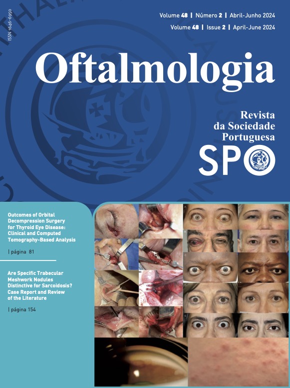
Outcomes of Orbital Decompression Surgery for Thyroid Eye Disease: Clinical and Computed Tomography-Based Analysis
DOI:
https://doi.org/10.48560/rspo.32537Keywords:
Decompression, Surgical, Graves Ophthalmopathy, Orbit/surgery, Tomography, X-Ray ComputedAbstract
INTRODUCTION: Orbital decompression surgery has been widely used in thyroid eye disease (TED). It is performed in the active stage, in sight-threatening cases of dysthyroid compressive optic neuropathy (DON), or severe corneal exposure unresponsive to steroids, and also during the quiescent phase to address proptosis. The purpose of this study is to analyze both clinical and imagiological outcomes of patients with TED who underwent orbital decompression.METHODS: A retrospective analysis of patients undergoing orbital decompression in Centro Hospitalar Universitário de Lisboa Central and Hospital Cuf Descobertas, between 2018 and 2021, was performed. Demographic and clinical data were collected. The procedures included lateral, inferior-medial, balanced (lateral and medial) and three-wall decompressions. Main clinical outcomes included best corrected visual acuity (BCVA), proptosis reduction and complications. A group of patients underwent orbital computed tomography (CT) scanning before and after surgery, and differences in globe displacement in the horizontal, vertical and anteroposterior planes were measured for each type of surgery.
RESULTS: Forty-six orbits from 28 patients (18 females and 10 males) underwent decompression surgery. Mean age at time of surgery was 49.43 ± 11.63 years old. Orbital decompression was performed during the inactive phase in 19 patients (67.4%) and was required to treat sight-threatening active TED in 9 patients (32.6%). Lateral, inferior-medial, balanced and three-wall decompression were carried out in 12 (26.1%), 8 (17.4%), 14 (30.4%) and 12 (26.1%) orbits, respectively. From baseline, statistically significant improvements were observed after surgery in logMAR BCVA (p<0.05) and proptosis (p<0.001). Larger proptosis reduction occurred in three-wall decompression, followed by balanced decompression (p<0.001). New-onset strabismus occurred in 3 of the 28 patients (10.7%): 1 endoscopic inferior-medial decompression, 1 three-wall decompression with an endoscopic approach to the inferior and medial walls and 1 transorbital three-wall decompression. All the 3 cases presented DON non-reversible with high dose intravenous steroids.
CONCLUSION: Orbital decompression is an effective procedure to address proptosis in TED, being also an important resource in cases of DON unresponsive to systemic steroids. The reduction in proptosis is associated with the number of orbital walls addressed. An individualized approach is crucial during surgical planning in TED cases.
Downloads
References
Bartalena L, Kahaly GJ, Baldeschi L, Dayan CM, Eckstein A, Marcocci C, et al. The 2021 European Group on Graves’ orbitopathy (EUGOGO) clinical practice guidelines for the medical management of Graves’ orbitopathy. Eur J Endocrinol. 2021;185:G43-67. doi:10.1530/EJE-21-0479.
Smith TJ. Pathogenesis of Graves’ orbitopathy: a 2010 update. J Endocrinol Invest. 2010;33:414-21. doi:10.1007/BF03346614.
Bartalena L, Piantanida E, Gallo D, Lai A, Tanda ML. Epidemiology, Natural History, Risk Factors, and Prevention of Graves’ Orbitopathy. Front Endocrinol. 2020;11:615993. doi:10.3389/fendo.2020.615993.
Fichter N, Guthoff RF, Schittkowski MP. Orbital decompression in thyroid eye disease. ISRN Ophthalmol. 2012;2012:739236. doi:10.5402/2012/739236
Choi SW, Lee JY, Lew H. Customized Orbital Decompression Surgery Combined with Eyelid Surgery or Strabismus Surgery in Mild to Moderate Thyroid-associated Ophthalmopathy. Korean J Ophthalmol. 2016;30:1-9. doi:10.3341/kjo.2016.30.1.1.
Goldberg RA, Perry JD, Hortaleza V, Tong JT. Strabismus after balanced medial plus lateral wall versus lateral wall only orbital decompression for dysthyroid orbitopathy. Ophthalmic Plast Reconstr Surg. 2000;16:271-7. doi:10.1097/00002341200007000-00004
Rocchi R, Lenzi R, Marinò M, Latrofa F, Nardi M, Piaggi P, et al. Rehabilitative orbital decompression for Graves’ orbitopathy: risk factors influencing the new onset of diplopia in primary gaze, outcome, and patients’ satisfaction. Thyroid. 2012;22:1170-5. doi:10.1089/thy.2012.0272
Sellari-Franceschini S, Lenzi R, Santoro A, Muscatello L, Rocchi R, Altea MA, et al. Lateral wall orbital decompression in Graves’ orbitopathy. Int J Oral Maxillofac Surg. 2010;39:16-20. doi:10.1016/j.ijom.2009.10.011
Mourits MP, Prummel MF, Wiersinga WM, Koornneef L. Clinical activity score as a guide in the management of patients with Graves’ ophthalmopathy [published correction appears in Clin Endocrinol.1997;47:632]. Clin Endocrinol. 1997;47:9-14. doi:10.1046/j.1365-2265.1997.2331047.x
Huh J, Park SJ, Lee JK. Measurement of proptosis using computed tomography based three-dimensional reconstruction software in patients with graves’ orbitopathy. Sci Rep. 2020;10:14554. doi: 10.1038/s41598-020-71098-4.
Dunsky IL. Normative data for Hertel exophthalmometry in a normal adult black population. Optom Vis Sci. 1992;69:562-4. doi:10.1097/00006324-199207000-00009.
Migliori ME, Gladstone GJ. Determination of the normal range of exophthalmometric values for black and white adults. Am J Ophthalmol. 1984;98:438-42. doi:10.1016/0002-9394(84)90127-2.
O’Donnell NP, Virdi M, Kemp EG. Hertel exophthalmometry: the most appropriate measuring technique. Br J Ophthalmol. 1999;83:1096B. doi:10.1136/bjo.83.9.1096b
Kim IT, Choi JB. Normal range of exophthalmos values on orbit computerized tomography in Koreans. Ophthalmologica. 2001;215:156-62. doi:10.1159/000050850.
Musch DC, Frueh BR, Landis JR. The reliability of Hertel exophthalmometry. Observer variation between physician and lay readers. Ophthalmology. 1985;92:1177-80. doi:10.1016/s0161-6420(85)33880-0
Lam AK, Lam CF, Leung WK, Hung PK. Intra-observer and inter-observer variation of Hertel exophthalmometry. Ophthalmic Physiol Opt. 2009;29:472-6. doi:10.1111/j.14751313.2008.00617.x
Kashkouli MB, Beigi B, Noorani MM, Nojoomi M. Hertel exophthalmometry: reliability and interobserver variation. Orbit. 2003;22:239-45. doi:10.1076/orbi.22.4.239.17245.
Frueh BR, Garber F, Grill R, Musch DC. Positional effects on exophthalmometer readings in Graves’ eye disease. Arch Ophthalmol. 1985;103:1355-6. doi:10.1001/archopht.1985.01050090107043
Ameri H, Fenton S. Comparison of unilateral and simultaneous bilateral measurement of the globe position, using the Hertel exophthalmometer. Ophthalmic Plast Reconstr Surg. 2004;20:448-51. doi:10.1097/01.iop.0000143712.42344.8c
Fichter N, Guthoff RF. Results after En Bloc Lateral Wall Decompression Surgery with Orbital Fat Resection in 111 Patients with Graves’ Orbitopathy. Int J Endocrinol. 2015;2015:860849. doi:10.1155/2015/860849
Genere N, Stan MN. Current and Emerging Treatment Strategies for Graves’ Orbitopathy. Drugs. 2019;79:109-24. doi:10.1007/s40265-018-1045-9
Boddu N, Jumani M, Wadhwa V, Bajaj G, Faas F. Not All Orbitopathy Is Graves’: Discussion of Cases and Review of Literature. Front Endocrinol. 2017;8:184. doi:10.3389/fendo.2017.00184
Müller-Forell W, Kahaly GJ. Neuroimaging of Graves’ orbitopathy. Best Pract Res Clin Endocrinol Metab. 2012;26:259-71. doi:10.1016/j.beem.2011.11.009
Ben Simon GJ, Syed HM, Lee S, Wang DY, Schwarcz RM, McCann J, et al. Strabismus after deep lateral wall orbital decompression in thyroid-related orbitopathy patients using automated hess screen [published correction appears in Ophthalmology. 2006;113:1622. Syed, Ahmad M [corrected to Syed, Hasan M]]. Ophthalmology. 2006;113:1050-5. doi:10.1016/j.ophtha.2006.02.015
Goldberg RA, Perry JD, Hortaleza V, Tong JT. Strabismus after balanced medial plus lateral wall versus lateral wall only orbital decompression for dysthyroid orbitopathy. Ophthalmic Plast Reconstr Surg. 2000;16:271-7. doi:10.1097/00002341200007000-00004
Rocchi R, Lenzi R, Marinò M, Latrofa F, Nardi M, Piaggi P, et al. Rehabilitative orbital decompression for Graves’ orbitopathy: risk factors influencing the new onset of diplopia in primary gaze, outcome, and patients’ satisfaction. Thyroid. 2012;22:1170-5. doi:10.1089/thy.2012.0272
Sellari-Franceschini S, Lenzi R, Santoro A, Muscatello L, Rocchi R, Altea MA, et al. Lateral wall orbital decompression in Graves’ orbitopathy. Int J Oral Maxillofac Surg. 2010;39:16-20. doi:10.1016/j.ijom.2009.10.011
European Group on Graves’ Orbitopathy (EUGOGO), Mourits MP, Bijl H, Altea MA, Baldeschi L, Boboridis K, et al. Outcome of orbital decompression for disfiguring proptosis in patients with Graves’ orbitopathy using various surgical procedures. Br J Ophthalmol. 2009;93:1518-23. doi:10.1136/bjo.2008.149302
Borumandi F, Hammer B, Kamer L, von Arx G. How predictable is exophthalmos reduction in Graves’ orbitopathy? A review of the literature. Br J Ophthalmol. 2011;95:1625-30. doi:10.1136/bjo.2010.181313
Leong SC, Karkos PD, Macewen CJ, White PS. A systematic review of outcomes following surgical decompression for dysthyroid orbitopathy. Laryngoscope. 2009;119:1106-15. doi:10.1002/lary.20213
Jefferis JM, Jones RK, Currie ZI, Tan JH, Salvi SM. Orbital decompression for thyroid eye disease: methods, outcomes, and complications. Eye. 2018;32:626-36. doi:10.1038/eye.2017.260
Choe CH, Cho RI, Elner VM. Comparison of lateral and medial orbital decompression for the treatment of compressive optic neuropathy in thyroid eye disease. Ophthalmic Plast Reconstr Surg. 2011;27:4-11. doi:10.1097/IOP.0b013e3181df6a87
Baril C, Pouliot D, Molgat Y. Optic neuropathy in thyroid eye disease: results of the balanced decompression technique. Can J Ophthalmol. 2014;49:162-6. doi:10.1016/j.jcjo.2013.10.006
Graham SM, Brown CL, Carter KD, Song A, Nerad JA. Medial and lateral orbital wall surgery for balanced decompression in thyroid eye disease. Laryngoscope. 2003;113:1206-9. doi:10.1097/00005537-200307000-00017
Sellari-Franceschini S, Berrettini S, Santoro A, Nardi M, Mazzeo S, Bartalena L, et al. Orbital decompression in graves’ ophthalmopathy by medial and lateral wall removal. Otolaryngol Head Neck Surg. 2005;133:185-9. doi:10.1016/j.otohns.2005.02.006
Unal M, Leri F, Konuk O, Hasanreisoğlu B. Balanced orbital decompression combined with fat removal in Graves ophthalmopathy: do we really need to remove the third wall? Ophthalmic Plast Reconstr Surg. 2003;19:112-8. doi:10.1097/01. IOP.0000056145.71641.F5
Shorr N, Neuhaus RW, Baylis HI. Ocular motility problems after orbital decompression for dysthyroid ophthalmopathy. Ophthalmology. 1982;89:323-8. doi:10.1016/s01616420(82)34793-4
Eing F, Abbud CM, Velasco e Cruz AA. Cosmetic orbital inferomedial decompression: quantifying the risk of diplopia associated with extraocular muscle dimensions. Ophthalmic Plast Reconstr Surg. 2012;28:204-7. doi:10.1097/IOP.0b013e31824dd8a0
Rootman DB, Golan S, Pavlovich P, Rootman J. Postoperative Changes in Strabismus, Ductions, Exophthalmometry, and Eyelid Retraction After Orbital Decompression for Thyroid Orbitopathy. Ophthalmic Plast Reconstr Surg. 2017;33:289-93. doi:10.1097/IOP.0000000000000758
Gomi CF, Yang SW, Granet DB, Kikkawa DO, Langham KA, Banuelos LR, et al. Change in proptosis following extraocular muscle surgery: effects of muscle recession in thyroid-associated orbitopathy. J AAPOS. 2007;11:377-80. doi:10.1016/j.jaapos.2007.01.115)
Ben Simon GJ, Mansury AM, Schwarcz RM, Lee S, McCann JD, Goldberg RA. Simultaneous orbital decompression and correction of upper eyelid retraction versus staged procedures in thyroid-related orbitopathy. Ophthalmology. 2005;112:92332. doi:10.1016/j.ophtha.2004.12.028
Lagrèze WA, Gerling J, Staubach F. Changes of the lid fissure after surgery on horizontal extraocular muscles. Am J Ophthalmol. 2005;140:1145-6. doi:10.1016/j.ajo.2005.06.060
Santos de Souza Lima LC, Velarde LG, Vianna RN, Herzog Neto G. The effect of horizontal strabismus surgery on the vertical palpebral fissure width. J AAPOS. 2011;15:473-5. doi:10.1016/j.jaapos.2011.05.017
Cruz AA, Equitério B, Diniz SB, Garcia DM, Rootman DB, Goldberg RA, et al. Upper Eyelid Contour Changes After Orbital Decompression in Graves Orbitopathy. Oph- thalmic Plast Reconstr Surg. 2022;38:289-93. doi: 10.1097/IOP.0000000000002093.
Ben Simon GJ, Mansury AM, Schwarcz RM, Lee S, McCann JD, Goldberg RA. Simultaneous orbital decompression and correction of upper eyelid retraction versus staged procedures in thyroid-related orbitopathy. Ophthalmology. 2005;112:92332. doi:10.1016/j.ophtha.2004.12.028
Downloads
Published
How to Cite
Issue
Section
License
Copyright (c) 2024 Revista Sociedade Portuguesa de Oftalmologia

This work is licensed under a Creative Commons Attribution-NonCommercial 4.0 International License.
Do not forget to download the Authorship responsibility statement/Authorization for Publication and Conflict of Interest.
The article can only be submitted with these two documents.
To obtain the Authorship responsibility statement/Authorization for Publication file, click here.
To obtain the Conflict of Interest file (ICMJE template), click here


