
Optimizing UV-A Irradiation Profiles in Crosslinking for Keratoconus: Comparison of Outcomes After Standard Accelerated and Topography-Guided Protocols
DOI:
https://doi.org/10.48560/rspo.33264Keywords:
Keratoconus, Crosslinking Reagents, Corneal Topography, Patient Outcome Assessment, Visual Acuity, Ultraviolet RaysAbstract
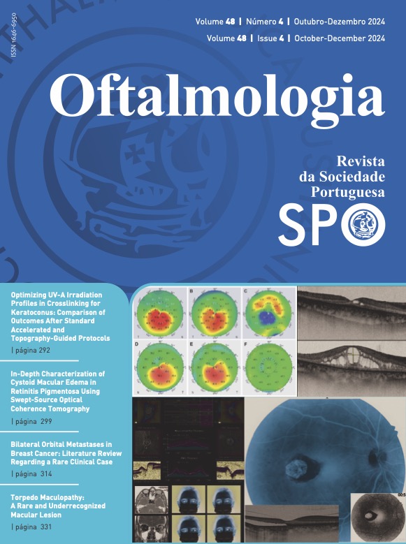
INTRODUCTION: Our purpose was to compare visual and tomographic outcomes of crosslinking treatment for progressive keratoconus, utilizing excimer-laser assisted epithelium removal and either central uniform irradiation (C-CXL) or customized, topography-guided irradiation (TG-CXL).METHODS: Retrospective study. We included patients with progressive keratoconus who underwent TG-CXL or C-CXL. In both procedures the epithelium was removed using phototherapeutic keratectomy (PTK) with a 50 µm ablation within a 7.0 mm optic zone, followed by riboflavin application every 2 minutes for 10 minutes. In TG-CXL this was followed by topography-guided ultraviolet-A (UVA) irradiation, with treatment energies ranging from 5.4 to 10 J/cm2 and fluence of 10 mw/cm2, while in C-CXL the cornea was uniformly irradiated with UVA with treatment energy of 6.0 J/cm2 and fluence of 10 mw/cm2. Patient data was collected at baseline, 6 and 12 months postoperatively including maximum keratometry (Kmax), anterior keratometry values within 5 points of a 3 mm diameter circle centered at the pupil, in the superior and inferior halves of the cornea, central corneal thickness, thinnest point pachymetry, subjective refraction and best corrected visual acuity (BCVA).
RESULTS: Fifty-four eyes from 48 patients were included (27 eyes for each group). Baseline characteristics were not significantly different between groups. Kmax was significantly lower 1 year (-0.83 ± 1.64 D; p=0.016) postoperatively for TG-CXL, but not for C-CXL (-0.46 ± 2.04 D; p=0.256). Inferior-superior (I-S) asymmetry index decreased significantly at 1 year for TG-CXL (-8.17 ± 9.56 D; p<0.001), but not for C-CXL (-3.69 ± 11.69 D; p=0.113). There were no significant differences in the evaluated structural parameters between 6 and 12 months postoperatively in both groups. The BCVA improved significantly at 1 year (difference to baseline: TG-CXL -0.13 ± 0.14 logMAR; p<0.001 and C-CXL -0.24 ± 0.38 logMAR; p=0.018; No difference between groups; p=0.244), while there was a significant myopic increase in spherical refractive error in both groups (difference to baseline: TG-CXL -1.05 ± 2.08 D; p=0.017 and C-CXL -0.90 ± 1.49 D; p=0.025).
CONCLUSION: One year after surgery, TG-CXL leads to greater Kmax reduction and topographic regularization than C-CXL. Both procedures lead to improved visual acuity and a myopic shift, with no superiority among the two. These results support the use of topography-guided crosslinking as a new valuable solution in the treatment of progressive keratoconus.
Downloads
References
Romero-Jiménez M, Santodomingo-Rubido J, Wolffsohn JS. Keratoconus: a review. Cont Lens Anterior Eye. 2010;33(4):157-166; quiz 205. doi:10.1016/j.clae.2010.04.006
Santodomingo-Rubido J, Carracedo G, Suzaki A, Villa-Collar C, Vincent SJ, Wolffsohn JS. Keratoconus: An updated review. Cont Lens Anterior Eye. 2022;45(3):101559. doi:10.1016/j.clae.2021.101559
Caporossi A, Mazzotta C, Baiocchi S, Caporossi T. Long-term results of riboflavin ultraviolet a corneal collagen cross-linking for keratoconus in Italy: the Siena eye cross study. Am J Ophthalmol. 2010;149(4):585-593. doi:10.1016/j.ajo.2009.10.021
Koller T, Mrochen M, Seiler T. Complication and failure rates after corneal crosslinking. J Cataract Refract Surg. 2009;35(8):1358-1362. doi:10.1016/j.jcrs.2009.03.035
Asri D, Touboul D, Fournié P, et al. Corneal collagen crosslinking in progressive keratoconus: multicenter results from the French National Reference Center for Keratoconus. J Cataract Refract Surg. 2011;37(12):2137-2143. doi:10.1016/j.jcrs.2011.08.026
Beckman KA, Gupta PK, Farid M, et al. Corneal crosslinking: Current protocols and clinical approach. J Cataract Refract Surg. 2019;45(11):1670-1679. doi:10.1016/j.jcrs.2019.06.027
Wu D, Lim DKA, Lim BXH, et al. Corneal Cross-Linking: The Evolution of Treatment for Corneal Diseases. Front Pharmacol. 2021;12:686630. doi:10.3389/fphar.2021.686630
Kymionis GD, Grentzelos MA, Kounis GA, Diakonis VF, Limnopoulou AN, Panagopoulou SI. Combined transepithelial phototherapeutic keratectomy and corneal collagen cross-linking for progressive keratoconus. Ophthalmology. 2012;119(9):1777-1784. doi:10.1016/j.ophtha.2012.03.038
Grentzelos MA, Liakopoulos DA, Siganos CS, Tsilimbaris MK, Pallikaris IG, Kymionis GD. Long-term Comparison of Combined t-PTK and CXL (Cretan Protocol) Versus CXL With Mechanical Epithelial Debridement for Keratoconus. J Refract Surg. 2019;35(10):650-655. doi:10.3928/1081597X-20190917-01
Roberts CJ, Dupps WJ. Biomechanics of corneal ectasia and biomechanical treatments. J Cataract Refract Surg. 2014;40(6):991-998. doi:10.1016/j.jcrs.2014.04.013
Nordström M, Schiller M, Fredriksson A, Behndig A. Refractive improvements and safety with topography-guided corneal crosslinking for keratoconus: 1-year results. Br J Ophthalmol. 2017;101(7):920-925. doi:10.1136/bjophthalmol-2016-309210
Cassagne M, Pierné K, Galiacy SD, Asfaux-Marfaing MP, Fournié P, Malecaze F. Customized Topography-Guided Corneal Collagen Cross-linking for Keratoconus. J Refract Surg. 2017;33(5):290-297. doi:10.3928/1081597X-20170201-02
Seiler TG, Fischinger I, Koller T, Zapp D, Frueh BE, Seiler T. Customized Corneal Cross-linking: One-Year Results. Am J Ophthalmol. 2016;166:14-21. doi:10.1016/j.ajo.2016.02.029
Shajari M, Steinwender G, Herrmann K, et al. Evaluation of keratoconus progression. Br J Ophthalmol. 2019;103(4):551-557. doi:10.1136/bjophthalmol-2017-311651
Mencucci R, Paladini I, Virgili G, Giacomelli G, Menchini U. Corneal thickness measurements using time-domain anterior segment OCT, ultrasound, and Scheimpflug tomographer pachymetry before and after corneal cross-linking for keratoconus. J Refract Surg. 2012;28(8):562-566. doi:10.3928/1081597X-20120703-02
Li Y, Tan O, Brass R, Weiss JL, Huang D. Corneal epithelial thickness mapping by Fourier-domain optical coherence tomography in normal and keratoconic eyes. Ophthalmology. 2012;119(12):2425-2433. doi:10.1016/j.ophtha.2012.06.023
Yang Y, Pavlatos E, Chamberlain W, Huang D, Li Y. Keratoconus detection using OCT corneal and epithelial thickness map parameters and patterns. J Cataract Refract Surg. 2021;47(6):759-766. doi:10.1097/j.jcrs.0000000000000498
Downloads
Published
How to Cite
Issue
Section
License
Copyright (c) 2024 Revista Sociedade Portuguesa de Oftalmologia

This work is licensed under a Creative Commons Attribution-NonCommercial 4.0 International License.
Do not forget to download the Authorship responsibility statement/Authorization for Publication and Conflict of Interest.
The article can only be submitted with these two documents.
To obtain the Authorship responsibility statement/Authorization for Publication file, click here.
To obtain the Conflict of Interest file (ICMJE template), click here

