
Optical Coherence Tomography (OCT) in Retinoblastoma Management: Experience of the Portuguese National Reference Center
DOI:
https://doi.org/10.48560/rspo.28264Keywords:
Disease Management, Retinoblastoma, Tomography, Optical CoherenceAbstract
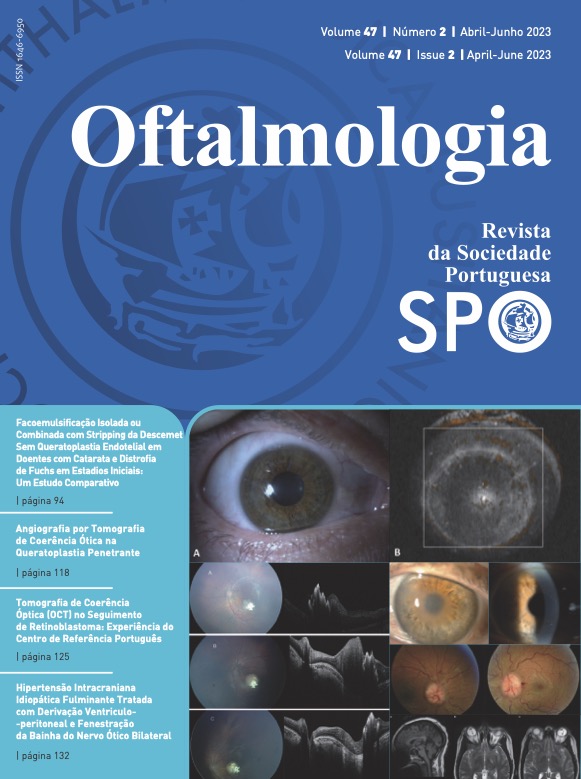
INTRODUCTION: Patients under 5 years old with retinoblastoma require close monitoring under anesthesia to ensure early detection of new tumors. They undergo monthly observations after diagnosis and during the first 6 months after treatment, then observations are gradually spaced in time until the age of five. Optical coherence tomography (OCT) is a relatively new exam in monitoring retinoblastoma patients. The advantages include pre-clinical diagnosis of new tumors when located in the posterior pole and evaluation of retinal layers in eyes submitted to intraarterial chemotherapy. We aimed to review the role of handheld OCT in evaluating eyes affected by retinoblastoma and to report our experience using this device.
METHODS: The observational case series included children with retinoblastoma followed at Centro Hospitalar e Universitário de Coimbra from January 2022 to August 2022 who underwent an OCT session during their routine observations under anesthesia. We collected data regarding patients’ age at presentation, family history, RB1 mutation status and International Intraocular Retinoblastoma Classification. The OCT images were analyzed for primary tumor characterization, relapse tumors and associated findings, and correlated to the fundus image on Retcam®.
RESULTS: We included 23 eyes of 19 children that had a total of 44 OCT exams. The median number of OCT scans per eye was 2 (range 1-6). The mean age at presentation of retinoblastoma was 9.37 months old. After reviewing the images, we were able to identify all 4 types of tumor remnants in our series. One patient had a new relapsed tumor that was detected primarily with OCT and treatment was initiated accordingly. In nine eyes, it was impossible to scan the primary tumor due to peripheral localization in the fundus due to advanced stages.
CONCLUSION: Hand-held OCT allows direct visualization of the retina and ensures a closer follow-up of young children with retinoblastoma, leading to the earlier diagnosis of relapses and the ability to treat them with less aggressive options, which may preserve more vision. The use and experience with OCT are increasing in all specialized centers that treat retinoblastoma. Hence, its usefulness will continue to grow and, in the future, there will be more clues to help diagnose retinoblastoma even sooner.
Downloads
References
Kivela T. The epidemiological challenge of the most frequent eye cancer: retinoblastoma, an issue of birth and death. Br J Ophthalmol. 2009;93:1129-31. doi: 10.1136/bjo.2008.150292
Jenkinson H. Retinoblastoma: diagnosis and management- the UK perspective. Arch Dis Child. 2015;100:1070-5. doi: 10.1136/archdischild-2014-306208
Yun J, Li Y, Xu CT, Pan BR. Epidemiology and Rb1 gene of retinoblastoma. Int J Ophthalmol. 2011;4:103-9. doi: 10.3980/j. issn.2222-3959.2011.01.24
Dimaras H, Corson TW, Cobrinik D, White A, Zhao J, Munier FL, et al. Retinoblastoma. Nat Rev Dis Primers. 2015;1:15021. doi: 10.1038/nrdp.2015.21
Medina CA, Plesec T, Singh AD. Optical coherence tomog- raphy imaging of ocular and periocular tumours. Br J Oph- thalmol. 2014;98 Suppl 2:ii40-46. doi: 10.1136/bjophthal- mol-2013-304299
Panozzo G, Parolini B, Gusson E, Mercanti A, Pinackatt S, Bertoldo G, et al. Diabetic macular edema: an OCT- based classification. Semin Ophthalmol. 2004;19:13-20. doi: 10.1080/08820530490519934
Quellec G, Lee K, Dolejsi M, Garvin MK, Abramoff MD, Sonka M. Three-dimensional analysis of retinal layer texture: identification of fluid-filled regions in SD-OCT of the macu- la. IEEE Trans Med Imaging. 2010;29:1321-30. doi: 10.1109/ TMI.2010.2047023
Scott AW, Farsiu S, Enyedi LB, Wallace DK, Toth CA. Imaging the infant retina with a hand-held spectral-domain optical co- herence tomography device. Am J Ophthalmol. 2009;147:364- 73 e362. doi: 10.1016/j.ajo.2008.08.010
Rootman DB, Gonzalez E, Mallipatna A, Vandenhoven C, Hampton L, Dimaras H, et al. Hand-held high-resolution spectral domain optical coherence tomography in retinoblas- toma: clinical and morphologic considerations. Br J Ophthalmol. 2013;97:59-65. doi: 10.1136/bjophthalmol-2012-302133
Samara WA, Pointdujour-Lim R, Say EA, Shields CL. Foveal microanatomy documented by SD-OCT following treatment of advanced retinoblastoma. J AAPOS. 2015;19:368-72. doi: 10.1016/j.jaapos.2015.02.019
Yousef YA, Shroff M, Halliday W, Gallie BL, Heon E. Detection of optic nerve disease in retinoblastoma by use of spectral domain optical coherence tomography. J AAPOS. 2012;16:481- 3. doi: 10.1016/j.jaapos.2012.05.010
Soliman SE, VandenHoven C, MacKeen LD, Gallie BL. Secondary Prevention of Retinoblastoma Revisited: Laser Pho- tocoagulation of Invisible New Retinoblastoma. Ophthalmol- ogy. 2020;127:122-7. doi: 10.1016/j.ophtha.2019.08.011
Nadiarnykh O, McNeill-Badalova NA, Gaillard MC, Bosscha MI, Fabius AW, Verbraak FD, et al. Optical coherence tomog- raphy (OCT) to image active and inactive retinoblastomas as well as retinomas. Acta Ophthalmol. 2020;98:158-65. doi: 10.1111/aos.14214
Soliman SE, VandenHoven C, MacKeen LD, Heon E, Gallie BL. Optical Coherence Tomography-Guided Decisions in Ret- inoblastoma Management. Ophthalmology. 2017;124:859-72. doi: 10.1016/j.ophtha.2017.01.052
Welch RJ, Rao R, Gordon PS, Say EAT, Shields CL. Optical Coherence Tomography of Small Retinoblastoma. Asia Pac J Ophthalmol. 2018;7:301-6. doi: 10.22608/APO.2018189
Linn Murphree A. Intraocular retinoblastoma: the case for a new group classification. Ophthalmol Clin North Am.
;18:41-53, viii. doi: 10.1016/j.ohc.2004.11.003
Shields CL, Palamar M, Sharma P, Ramasubramanian A, Leahey A, Meadows AT,et al. Retinoblastoma regression pat- terns following chemoreduction and adjuvant therapy in 557 tumors. Arch Ophthalmol. 2009;127:282-90. doi: 10.1001/archophthalmol.2008.626
Castela G, Providencia J, Monteiro M, Silva S, Brito M, Sá J, et
al. Characterization of the Portuguese population diagnosed with retinoblastoma. Sci Rep. 2022;12:4378. doi: 10.1038/ s41598-022-08326-6
Rothschild PR, Levy D, Savignoni A, Lumbroso-Le Rouic L, Aerts I, Gauthier-Villars M, et al. Familial retinoblastoma: fundus screening schedule impact and guideline proposal. A retrospective study. Eye. 2011;25:1555-61. doi: 10.1038/ eye.2011.198
Maldonado RS, Izatt JA, Sarin N, Wallace DK, Freedman S, Cotten CM, et al. Optimizing hand-held spectral domain optical coherence tomography imaging for neonates, infants, and children. Invest Ophthalmol Vis Sci. 2010;51:2678-85. doi: 10.1167/iovs.09-4403
Downloads
Published
How to Cite
Issue
Section
License
Copyright (c) 2023 Revista Sociedade Portuguesa de Oftalmologia

This work is licensed under a Creative Commons Attribution-NonCommercial 4.0 International License.
Do not forget to download the Authorship responsibility statement/Authorization for Publication and Conflict of Interest.
The article can only be submitted with these two documents.
To obtain the Authorship responsibility statement/Authorization for Publication file, click here.
To obtain the Conflict of Interest file (ICMJE template), click here

